Anti lamin a c antibody e 1 is a mouse monoclonal igg 1 kappa light chain lamin a c antibody provided at 200 µg ml.
Anti lamin a c antibody.
Raised against porcine lamin preparation.
Performed under reducing conditions.
Lamins are a class of intermediate filament proteins that form a matrix on the inner surface of the nuclear envelope.
70 kda ab8984 was used at a 1 100 dilution against human fibroblast lysate.
Western blot analysis of extracts of nci h293 cells using anti lamin a c antibody a0464.
Goat anti rabbit igg h l hrp as014 at 1 10 000 dilution.
Lamin a and c are detected.
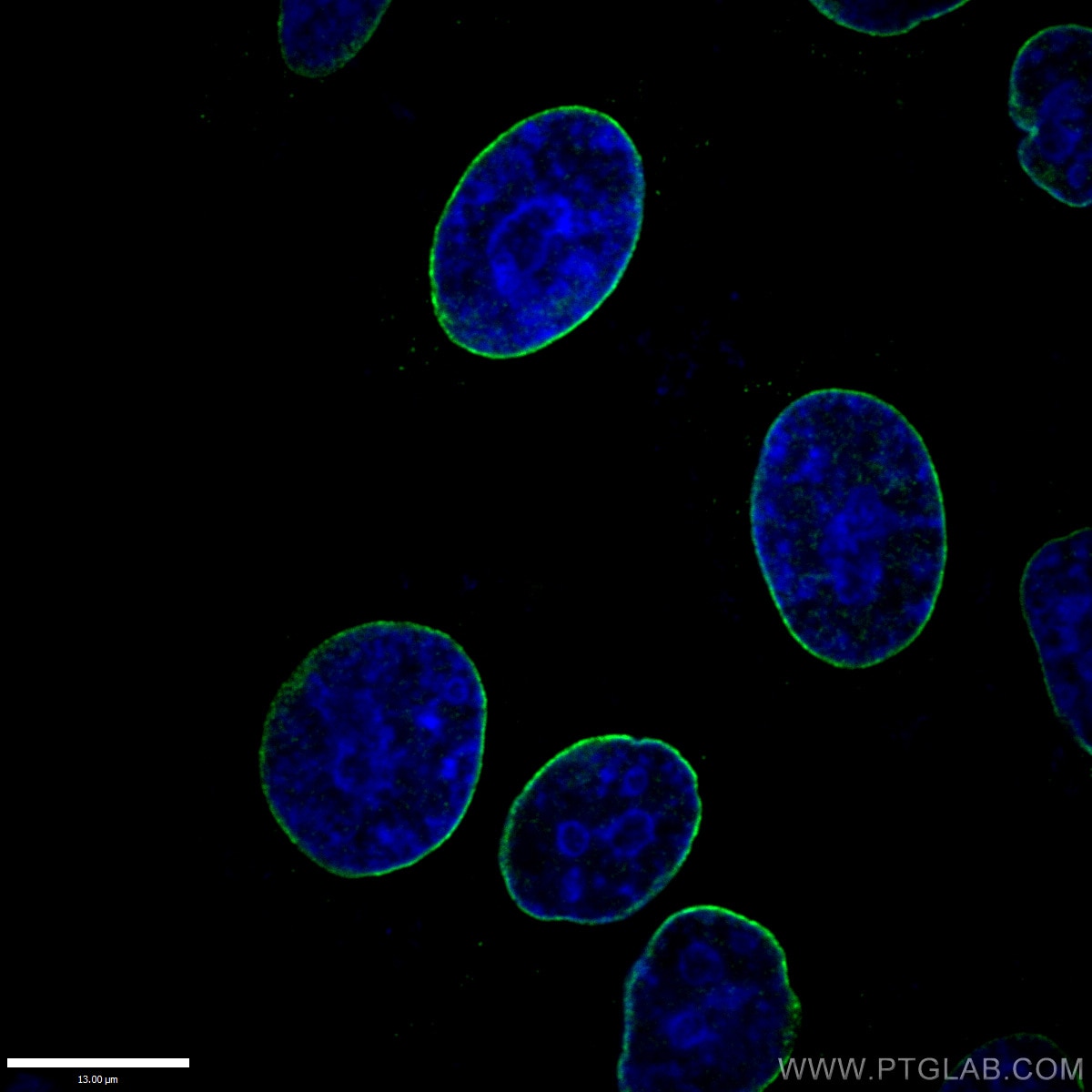
The anti lamin a c antibody reveals strong nuclear lamina staining while anti lamp1 antibody reveals strong cytoplasmic punctate staining of lysosomes and early endosomes.
Anti lamin a lamin c antibody 131c3 nuclear envelope marker ab8984 at 1 100 dilution human fibroblast cell lysate at 15 µl developed using the ecl technique.
Peptide from human lamin a c corresponding to amino acids 398 to 490.
These proteins are found in many different cell types in three different forms a b and c.
Specific for an epitope mapping between amino acids 2 29 at the n terminus of lamin a c of human origin.
Reacts against lamin a exclusively the antibody was raised against the carboxy terminus of 98 amino acids present in lamin a and absent from lamin c.
Since both dna blue and lamin a c red are associated with the nuclear compartment this region appears crimson in this image.
This antibody has been reported by an independent laboratory to detect lamin a c in human endothelial cells.
In humans this protein is encoded by the gene lmna.
See machiels et al 1997.
Anti lamin a c antibody 636 is recommended for detection of lamin a and lamin c of mouse rat and human origin by wb ip if ihc p and fcm.
Species predicted to react based on 100 sequence homology.
The expected protein mass is 74 1 kda but there are 6 reported isoforms.
Lamins a and c are alternatively spliced versions of the lmna gene.
The protein may also be known as lamin c dhe lamin a cdcd1 cddc and cmd1a.
Lamin a c antibodies.
As a result this antibody recognizes lamin a but not lamin c.
Anti lamin a c antibodies are available from several suppliers.